|
| Guess What! - ESTP Case 31 |
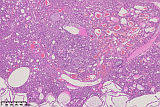
The lesion was observed as an incidental finding in the testis of a 22 months old male Wistar rat, which was found in a moribund condition after continued weight loss. At necropsy, one testis showed a glassy appearance.
| Click on the images below for a larger view. |
|
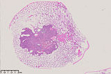
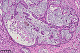
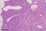
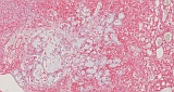
Fig. 1: H&E, 1x
|
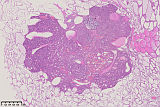
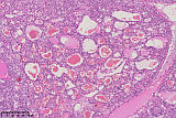
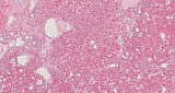
Fig. 2: H&E, 2x
|
|
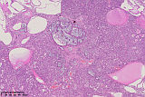
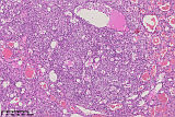
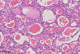
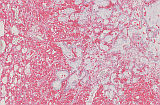
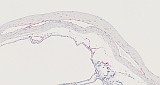
Fig. 3: H&E, 5x
|
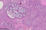
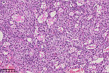
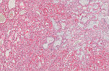
Fig. 4: H&E, 10x
|
|
Fig. 5: H&E, 20x
|
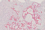
Fig. 6: H&E, 5x
|
|
Fig. 7: H&E, 10x
|
Fig. 8: H&E, 20x
|
|
Fig. 9: H&E, 5x
|
Fig. 10: H&E, 10x
|
|
Fig. 11: H&E, 20x
|
For case 31, we received 17 submissions reflecting the diagnostic difficulties of this case.
At first posting, we provided 11 images of an H&E slide. Thereupon we received 11 responses:
- Interstitial (Leydig) cell tumor.
- Rete testis hyperplasia in progression to adenoma. Seminiferous tubule degeneration/atrophy marked diffuse.
- Benign teratoma.
- Leydig cell adenoma (retiform).
- In the testis, we can see an advanced atrophy of the seminiferous tubules (age related). In the center of the testis we observed an interstitial cell tumor, associated with a glandular hyperplasia.
- Leydig cell adenoma, benign. Consisted of foamy to vacuolated cytoplasm containing Leydig cells. Expansion is more than 3 SFT and atrophy of rest of SFT. Cyst-like structure is present containing proteinaceous fluid and blood cells. No prominent compression observed. Fibrosis and dilatation of rete tubule seen.
- Bronchiolo-alveolar adenoma with marked infiltration of foamy macrophages and blood aspiration.
- Not an easy one... there seems to be a lot of foamy cells which are the majority of the tumor mass and resemble Leydig cells. Yet the tubular structures are strange....so this makes me thinking of a mixed form?!
- Carcinoma of the rete testis. Don't see packeting appearance for Sertoli cell tumor. Should this be vimentin negative? Could CK7 IHC use to differentiate that from Sertoli?
- Morphology consists with Interstitial cell tumor, in which the edema, dilation and congestion of vascular might cause the glassy appearance. Focally, there is ductal structures that look like epididymis ductal metaplasia.
- It's a tumor within the testicle. The tumor is richly vascularized and infiltrated with several types of inflammatory cells, mainly foamy poplar macrophages, and is considered to be a metastatic tumor of the lymphatic system.
| Click on the images below for a larger view. |
|
Fig. 12: Pan-Cytokeratin, 10x
|
Fig. 13: Vimentin, 10x
|
|
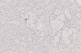
Fig. 14: S-100, 10x
|
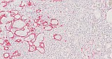
Fig. 15: Podoplanin, 10x
|
To further characterize the lesion, we performed immunohistochemistry with antibodies against vimentin, pan-cytokeratin, S-100, and podoplanin. We have posted a photo for every label. After that, we received 6 additional diagnoses.
- Something linked to embryonal gestation, e.g. a yolk sac tumor or a mixed sex cord germ cell tumor.
- Leydig cell adenoma.
- Majority of the tumor cells are arranged in packets with S-100 and Vimentin positivity, indicating its neuroendocrine origin, mixed with the presence of well differentiated glands containing mucinous materials and Pan-CK positivity of the epithelia, and some sinusoid structures with erythrocytes, suggested this tumor contains multiple components from endoderm, exoderm and mesoderm.
- Testes, Teratoma.
- Adenomatoid tumor in testes.
- Gonadoblastoma.
Description of the case
The introductory report of macroscopic observations, which was posted together with the first photos, suggested a lesion of a testis of a male Wistar rat.
The overview photograph and low magnification show a dense mass surrounded by mostly round degenerated or atrophic seminiferous tubules. 16 of 17 senders diagnosed or suspected a tumor in the testis.
The lesion is located in the middle of the testis section with compression to the surrounding tubules, but without invasion to the testis capsule or adjacent tissue.
Figures 3 to 5, 6 to 8 and 9 to 11 show low, medium, and high magnification, respectively, of lesion-specific features within the tumor-suspicious area.
Figures 3 to 5 show a glandular structure consisting of cuboidal cells surrounding a lumen containing a blue mucous-like substance. Around the glandular structures there are areas of fibrous cells of the tumor stroma.
Figures 6 to 8 show large uniform polyhedral cells with small round central nuclei and eosinophilic cytoplasm, which is mostly finely granular or vacuolated. Interspersed with these cells are several tubular lumens and a few vessels. The nuclear to cytoplasm ratio is low. There is no cellular polymorphism or atypia. Mitotic activity is low, there are no atypical mitotic figures. In this area of large vacuolated polyhedral cells, the tumor stroma is sparse.
Figures 9 to 11 show an area of several vessels and tubular structures with vacuolated cells between them.
| Click on the images below for a larger view. |
|
Fig. 16: Pan-Cytokeratin, 20x
|
Fig. 17: Vimentin, 10x
|
|
Fig. 18: S-100, 10x
|
Fig. 19: Podoplanin, 20x
|
Immunohistochemistry of the case
Pan-Cytokeratin:
Pan-cytokeratin is mainly a marker for epithelial cells. Some cytokeratin types are expressed by Seminomas, in developing Sertoli cells, but none by Leydig cells. Cytokeratins are present in the rete testis.
The tubular structures of the tumor show a strong expression for pan-cytokeratin, but all other parts of the tumor are negative (see Fig. 16).
Vimentin:
Vimentin is a cytoskeletal protein expressed mainly in cells of mesenchymal origin. Therefore, it is present in fibroblasts, endothelial cells, and cells of the immune system. Vimentin is expressed by both Sertoli and Leydig cells, and tumors arising from these cells, but not seminomas.
All parts of the tumor, except the tubular structures were positive for vimentin (see Fig. 17).
S-100:
S-100, a protein originally believed to be unique to the nervous system, has been found in extraneural presence of S-100 in the testis, namely in Leydig cells. In addition, S-100 proteins are normally present in cells derived from the neural crest (Schwann cells, and melanocytes), chondrocytes, adipocytes, myoepithelial cells, macrophages, Langerhans cells, dendritic cells, and keratinocytes.
The vacuolated tumor cells show a strong expression for S-100, the tubular structures stain weakly positive (see Fig. 18).
Podoplanin:
Podoplanin is a marker for lymphoid epithelial cells and as well for mesothelial cells (e.g. mesotheliomas of the epithelioid or biphasic type) (see Fig. 19).
The tumor is completely negative except of some superficial lymph vessels which are positive for podoplanin.
Proposed Diagnosis
- Adenoma, Leydig cell: Leydig cell adenoma consists of cells as seen here in this lesion: predominantly of uniform polyhedral cells with abundant eosinophilic, finely granular, or vacuolated cytoplasm. Unusual is the occurrence of epithelial-like structures with such tumors except some included rete testis structures. Leydig cell adenomas in rats may show a "Retiform pattern". In INHAND the features are specified as follows: Leydig cell tumor with embedded areas of glandular/tubular structures, lined by cuboidal to columnar cells with Alcian blue-positive brush borders, occasionally filled with PAS-positive substance.
Because of the histological features of the tumor of case 31 and the immunohistochemistry supplied to exclude other tumor types, we propose the diagnosis of an "Leydig cell adenoma, with retiform pattern". Such tumors with exaggerated retiform proliferations are rare events.
Discussion of Differential diagnoses
- Carcinoma, Leydig cell: Polymorphism, cellular atypia, and invasion of adjacent tissue (vessels, capsule) or formation of metastases should be present, which is not the case here.
- Adenoma, rete testis/Carcinoma, rete testis: A rete testis adenoma consists of tubular-papillary structures similar to the presented tumor but does not contain other cell types such as those present in this case. In the current lesion the tubular structures are surrounded by proliferating vacuolated cells. A rete testis carcinoma shows in addition presence of malignant characteristics such as invasion, marked atypia, high mitotic rate, and hemorrhage, which is not the case here.
- Hyperplasia, rete testis: Architecture of the rete testis should be maintained, which is not the case here.
- Teratoma, benign/malignant: Teratomas must contain tissue derived from the three germ layers. Tissue components are generally well differentiated. Benign teratomas often contain cysts lined by cuboidal, enteric, or respiratory epithelium. Smooth muscle may surround these cysts. Shows peripheral invasion or metastases. In general, the differential diagnosis must be discussed. At the first glance, the positivity for S-100, pan-cytokeratin and vimentin suggest this diagnosis, but knowing that S-100 is expressed by Leydig cells the immunohistochemistry pattern appears in another light and leads together with the histological appearance of the granular or vacuolated cells to the diagnosis of a tumor containing Leydig cells.
- Carcinoma, embryonal: Composed of large and anaplastic epithelium-like cells with a primitive appearance and indistinct cell boundaries. The cells do not have an anaplastic appearance and the cell boundaries in this case are clear. Nuclei are large with coarse chromatin and prominent nucleoli. In the present case the nuclei are small. Tumor cells may occur in solid sheets or have interspersed areas of acinar, papillary, or tubular structures. Intratubular growth is considered to represent an early tumor stage. Foci of yolk sac differentiation, choriocarcinoma, or well-differentiated tissues such as cartilage, bone, or skin may occur, this is not seen here.
- Gonadoblastoma: A gonadoblastoma is a complex neoplasm composed of a mixture of gonadal elements, such as large primordial germ cells, immature Sertoli cells or granulosa cells of the sex cord, and gonadal stromal cells. Gonadoblastomas in humans are, by definition, benign. Blastomas generally contain predominantly cells with basophilic cytoplasm, in the presented tumor the cytoplasm is more eosinophilic.
- Mesothelioma, malignant: Epithelioid mesothelioma or biphasic mesothelioma may contain epithelioid poorly formed glandular structures or ill-formed glands. In the testis region, mesothelioma occasionally arises at the tunica vaginalis and shows frequently a spread on the testis surface. To exclude a mesothelioma, anti-podoplanin was applied with a negative result in the lesion.
- Yolk sac tumor: Carcinoma, Yolk sac – Most characteristic features are tumor cells producing abundant eosinophilic, PAS-positive matrix in which neoplastic cells are embedded. Because there is no typical matrix-like structure, a PAS stain was not performed. Cell types and patterns that mimic the two layers of fetal membranes, i.e., the parietal and visceral yolk sac, are not observed here. Visceral yolk sac endodermal cells do not contain eosinophilic, PAS-positive droplets and may be columnar or large polygonal cells with pale, vacuolated cytoplasm, and giant nuclei. No giant nuclei are observed in the presented tumor. Cell nucleus is dark with closely clumped chromatin and contains one or two inconspicuous nucleoli. The nucleus to cytoplasm ratio approaches 1:1. In the present case, the nuclear to cytoplasm ratio is low. The tumor spreads aggressively to peritoneal surfaces and grows as nests or large colonies but seldom invades into the parenchyma of solid organs. In the present case, there is no spread on the surface and no aggressive growth.
- Mixed sex cord germ cell tumor: In Sertoli-cell tumors, which are sex-cord stromal tumors, the cells are arranged in palisades on a delicate fibrovascular stroma resulting in the formation of tortuous tubular structures without distinct lumina. Tumor cells are elongated (here they are polygonal) with generally pale and vacuolated cytoplasm and finely stippled nuclear chromatin, cell borders indistinct (here cell borders are clearly visible).
- Benign granulosa cell tumor: Growth pattern reminiscent of ovarian granulosa cell tumors, e.g., forming follicle-like nests and nodule. Small, basophilic cells like those of ovarian granulosa cell tumors with scant eosinophilic cytoplasm and nuclei with single nucleolus and stippled chromatin. Cells are usually round but may be spindle-shaped or pleomorphic: Mitotic figures occasionally present.
In the present tumor, in contrast to granulosa cell tumors, most cells show abundant eosinophilic cytoplasm, which is finely granulated or vacuolated.
- Histiocytic sarcoma (the tumor was described to show foamy popular macrophages and was suspected to be a metastatic tumor of the lymphatic system): Histiocytic sarcoma may occur in the testis. It consists of uniform population of round or oval cells with abundant eosinophilic cytoplasm. Cells may also be spindle shaped. Nuclei are round, irregular, elongated, folded, or indented; ring-shaped nuclei with a central opening may occasionally be present. Multinucleate giant cells are often scattered throughout the tumor, which is not the case here. Histiocytic sarcomas are rarely positive for S-100 and do not contain tubular pan-cytokeratin-positive structures. The cells often remind to macrophages but do not show distinct cell borders like in our case. Therefore, it is an important differential diagnosis to Leydig-cell tumor but can be excluded by H&E appearance.
- Benign granular cell tumor: Granular cell tumors are circumscribed, well-demarcated solid masses composed of large round to oval cells with pale basophilic nuclei and abundant eosinophilic granular cytoplasm and smaller cells with small dark uniform nuclei. They usually show prominent interstitial collagen (which is not the case in the present tumor). Cytoplasmic granules react immunohisto-chemically positive with antibodies directed against S-100 and vimentin. The granules are considered to be different stages of lysosomes. The cell borders of granular cell tumors are somewhat indistinct, while the cell borders of Leydig cell tumor cells are clearly visible. The cytoplasm of granular cell tumor cells has fine granules but is not vacuolated. Leydig cell tumors can also have finely granulated cytoplasm, and usually also show fine vacuoles in the tumor cells.
Many thanks to all those who participated in submitting diagnoses and considerations and who suggested many different diagnoses, most of which represent important differential diagnoses. Our diagnosis suggestion is based on discussions, as the tumor is not so easy to diagnose. Such cases are relatively rare, but an example image (Fig 3, 4, and 7) of a similar tumor with ductular structures is shown with the INHAND goRENI term description.
This was the last case in the old format. Be prepared to welcome The "New Guess What" - Coming SOON.
References
- Alison R, Ettlin RA, Foley GI, Harleman JH, Maekawa A, McConnell RF, Quereshi SR, Rehm S, Reznik G, Valerio M (1997). In: Mohr U, Capen CC, Dungworth DL, Griesemer RA, Ito N, Turusov US (eds) International Classification of Rodent Tumours, The Rat male genital system, Part I, IARC Scientific Publication, No. 122, Lyon, France.
- Creasy D, Bube A, de Rijk E, Kandori H, Kuwahara M, Masson R, Nolte T, Reams R, Regan K, Rehm S, Rogerson P, Whitney K (2012) Proliferative and nonproliferative lesions of the rat and mouse male reproductive system. Toxicol Pathol 40: 40S–121S
- Dixon D, Alison R, Bach U, Colman K, Foley GL, Harleman JH, Haworth R, Herbert R, Heuser A, Long G, Mirsky M, Regan K, Van Esch E, Westwood FR, Vidal J, Yoshida M (2014) Nonproliferative and proliferative lesions of the rat and mouse female reproductive system. J Toxicol Pathol 27: 1S–107S
- Emerson RE, Ulbright TM (2005) The use of immunohistochemistry in the differential diagnosis of tumors of the testis and paratestis. Semin Diagn Pathol 22: 33–50
- Ordóñez NG (2005) D2-40 and podoplanin are highly specific and sensitive immunohistochemical markers of epithelioid malignant mesothelioma. Hum Pathol 36: 372–380
- Qureshi SR, Perentes E, Ettlin RA, Kolopp M, Prentice DE, Frankfurter A (1991) Morphologic and immunohistochemical characterization of Leydig cell tumor variants in Wistar rats. Toxicol Pathol 19: 280–286
|
|